The CT scan is more useful for identification of blood clots in the lung. Although blood clots are harmful they can be treated.
Doing this then allows for the blood clot to show prominently on an X-ray.

Can you see a blood clot on an x ray. When paired with other symptoms excessive sweating can be one of the blood clot symptoms you shouldnt ignore located in either the lung or heart per the ASH. If you see any of the below-mentioned signs or symptoms it indicates that you would need to contact your health care practitioner for a checkup at the earliest. Blood clots in the stomach or an abdominal blood clot are a type of deep vein thrombosis DVT.
Perhaps one of the first and obvious evidences is pain which replies. Magnetic Resonance Imaging MRI An MRI can provide images of. This is a type of X-ray that gives a very detailed look at the leg veins in cross section and can detect clots.
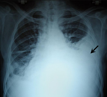
Next a medical professional takes radiographs or x-rays of the area where doctors suspect blood clots. A chest x-ray cannot prove that PE is present or absent because clots do not show up on x-ray. Symptoms Of Blood Clots.
CT scans looking for blood clots also use dye that can damage the kidneys or cause an allergic reaction. It is rarely used for this purpose as it is more difficult to interpret and is time consuming. This imaging test may be used to rule out blood clots in parts of your body other than your arm.
Do you know that there is blood clot in toe even. Very rarely a chest x-ray can show typical signs of plumonary emboli. CT scans usually result in exposure to large doses of radiation which can increase the risk of cancer over your lifetime.
Chest-x-ray is performed to rule out other causes of symptoms such as pneumonia or pleural effusion. Occasionally when pulmonary infarction occurs the x-ray may suggest this diagnosis although more testing is necessary. Nevertheless a chest x-ray is a useful test in the evaluation for PE because it can find other diseases such as pneumonia or fluid in the lungs that may explain a persons symptoms.
In order for blood clots to show on a radiograph contrast dye is injected into the body and the dye is absorbed by the blood clot. In some cases they may be a warning sign of an undiagnosed cancer but. If you notice the problems with it and its toes such as redness and swelling you should exclude a clot visiting a doctor.
A doctor will then perform an X-ray video X-ray of the affected area. It uses computers and X-rays to take cross-sectional images of your body. Pulmonary angiography can be used to diagnose a pulmonary embolus whereas venography is used to.
Hence you need to approach your doctor immediately once you begin to see any signs or symptoms. For these reasons if your risk of having a PE is low then the potential risks of a CT scan can outweigh the benefits. Angiography or Venography.
A radioactive dye contrast agent is injected into the top of your foot or ankle. You should be aware of any strange and new changes happening with your feet. The visual result will help a doctor to make a diagnosis.
An x ray will not show a blood clot in the lungs only a CT angiogram will show that. Early signs of blood clot in toe picture 3 can be difficult to interpret correctly. These are catheterization techniques in which a dye is injected into a blood vessel where a clot is suspected and X-rays are taken to detect the clot.
A doctor will be able to determine the location of the clot and also see decreased flow of blood in the blood vessels. An X-ray is then taken that enables your doctor to see images of your veins in your legs and feet so they can look for blood clots. Unlike traditional radiography which uses a single x-ray source that bounces off a detector plate to create an image CT scanning uses a chamber with multiple x-ray sources and detectors that spin around the body at tremendous velocity.
Ct with contrast agent or vq lung scan with radioisotopes inhalation and intravenous injection are the most common methods for diagnosing pulmonary emboli. Normally no they will not.
 A Case Of An Airway Obstruction Secondary To Blood Clot Formation After An Episode Of Massive Hemoptysis In A Smear Positive Pulmonary Tuberculosis Pregnant Lady Sciencedirect
 Organized Blood Clot Masquerading As Endobronchial Tumor A Review Of Management And Recent Advances Sciencedirect
 Copd X Ray Pictures Diagnosis And More
 7 Warning Signs Of A Pulmonary Embolism The Healthy
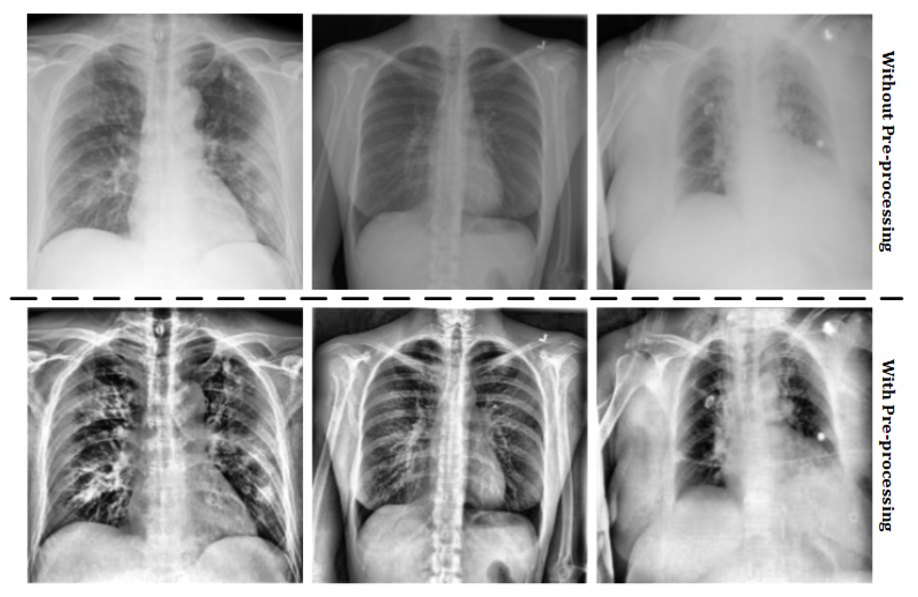 Using Lung X Rays To Diagnose Covid 19 Imaging Technology News
 Hd Chest X Ray In Pulmonary Embolism Em Rap
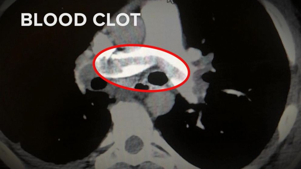 Why Are So Many Covid 19 Patients Also Seeing Blood Clots Abc News
 The Link Between Lung Cancer And Blood Clots Everyday Health
 Medical Notes Pulmonary Embolism Bbc News
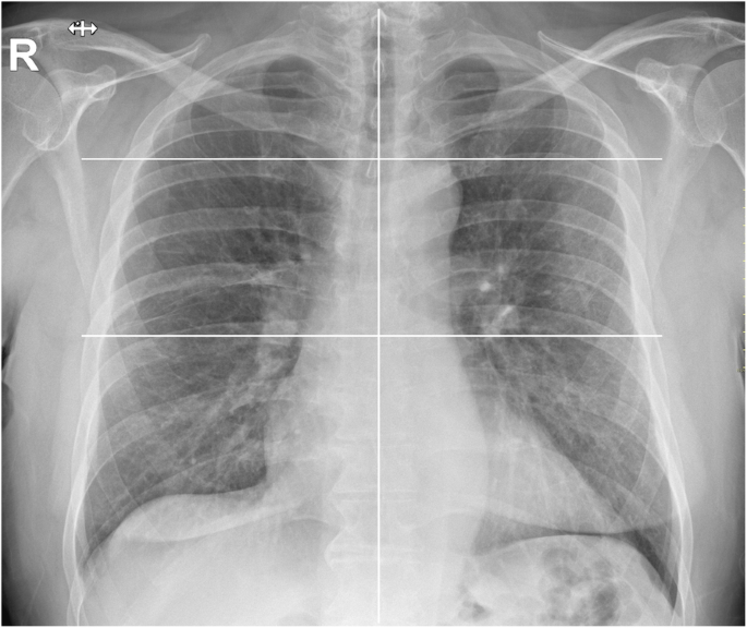 Chest X Ray Severity Score In Covid 19 Patients On Emergency Department Admission A Two Centre Study European Radiology Experimental Full Text
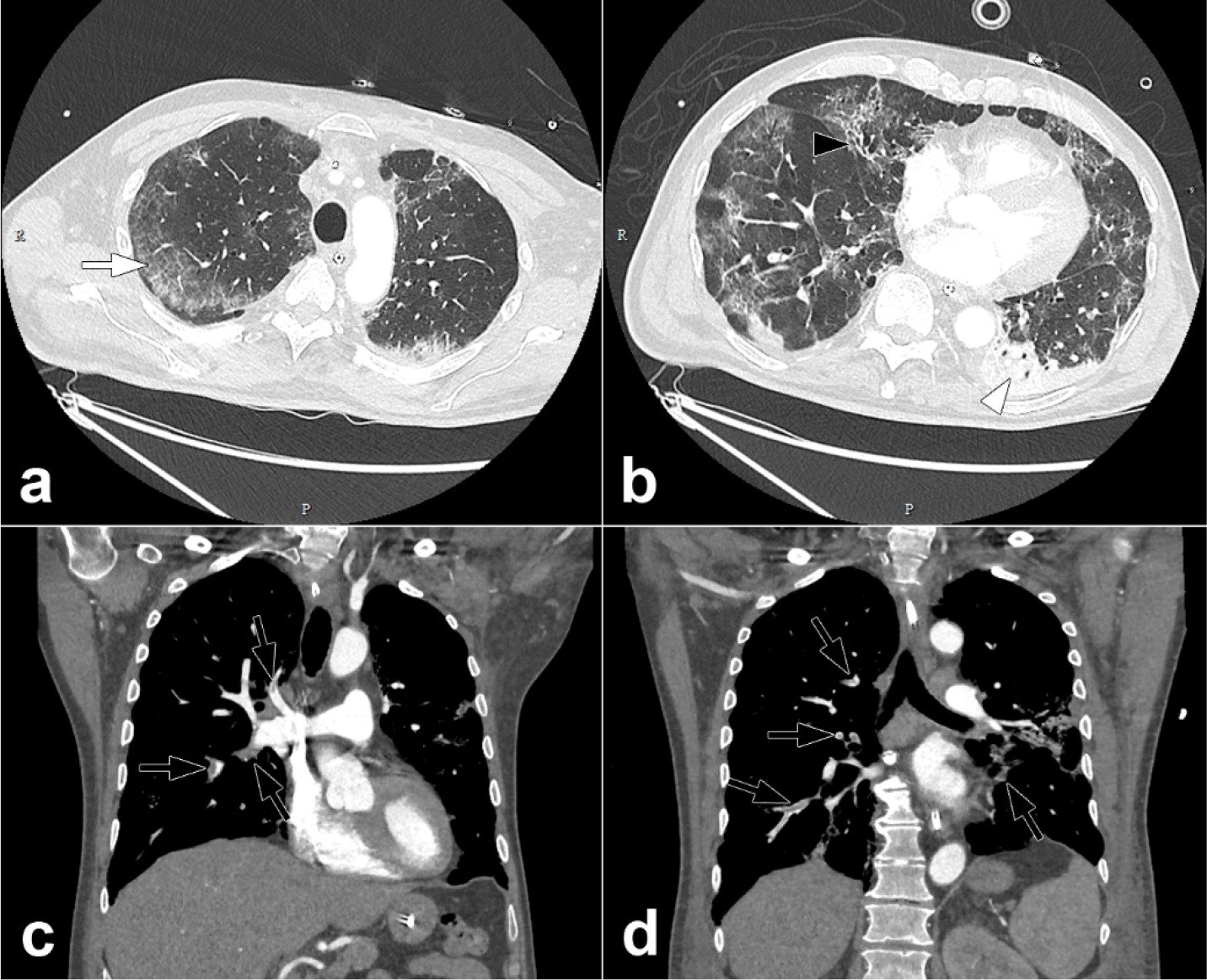 New Research Highlights Blood Clot Dangers Of Covid 19 Imaging Technology News
 Emergency Removal Of Sludged Blood From Main Bronchus With Cryotherapy Through Bronchoscope In Rescuing An Acute Respiratory Failure Caused By Massive Blood Clot Obstruction
 Suburban Imaging Understanding Blood Clots
 Chest X Rays For Recovered Covid 19 Patient In Grove City Revealed Blockage Blood Clots Wsyx
Tidak ada komentar:
Posting Komentar
Catatan: Hanya anggota dari blog ini yang dapat mengirim komentar.