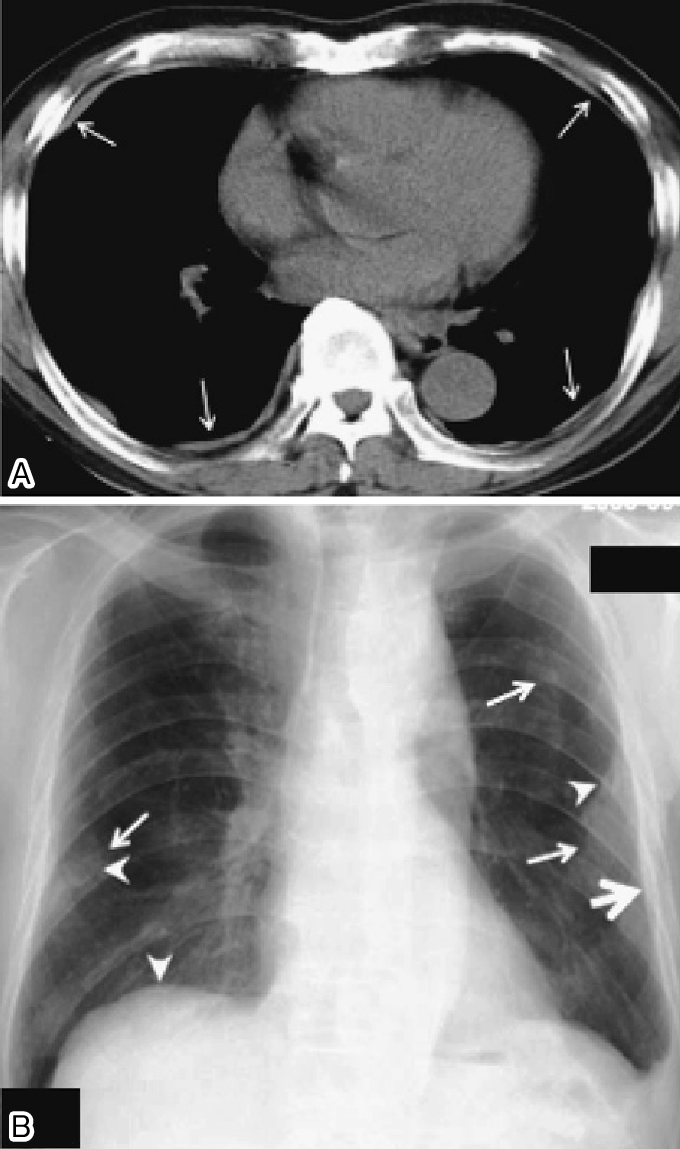
Pleural plaques are evidence of past exposure to asbestos and are the most common form of asbestos disease. Pleural plaques are the most common sign of past exposure to asbestos.
 Medpix Case Asbestos Related Pleural Plaques
Pleural plaques are the most common sign of asbestos exposure.
Asbestos pleural plaques. Pleural plaque is an indicator of asbestos exposure. Pleural plaques are sometimes referred to as hyaline pleural plaques. They are caused by exposure to asbestos but do not usually develop until some 20 40 years after first exposure.
Pleural plaques are fibrous or partially calcified scarring formed on the pleura caused by asbestos fibres. The relationship between a radiological semiquantitative score of pleural plaques and indices of asbestos exposure was also. The study population consisted of 2743 subjects presenting with no parenchymal interstitial abnormalities on the high-resolution.
The exposure may be occupational or environmental. Pleural plaques are essentially scars affecting the lungs and pleural membranes. Pleural plaques are areas of thickened tissue that form in the lining of the lung the pleura.
These deposits usually are found on the parietal pleura the membrane that lines the chest wall. Diagnosis of pleural plaques generally happens 20 to 40 years after initial asbestos exposure. This is because they are composed of cartilage-like tissue hyaline collagen.
The pleura is a two layered serous membrane lining surrounding both lungs that is attached to the internal chest wall. In those patients with bilateral pleural plaques detected radiographically an exposure history can be identified in 80 percent of patients. They are areas of slight fibrous thickening on the pleura the lining of the lungs and rib cage.
Although asbestos dust is not the sole cause of pleural plaques it is certainly the most common. Researchers believe that asbestos exposure is the main cause of pleural plaques. In most cases pleural plaques will develop only after prolonged asbestos exposure.
As with many asbestos diseases there is. Pleural plaques may take 10 to 30 years to be diagnosed and usually do not require treatment. These areas are called pleural plaques.
It is not however a form of cancer and should not be assumed that patients will later develop more serious diseases. Its opposite the visceral pleura wraps around the lungs. The commonest asbestos-related diseases are benign diseases and many studies have examined the relationships between asbestos exposure and these diseases 1Overall the prevalence of both pleural plaques and asbestosis is associated with time since first exposure TSFE to asbestos.
Chest pain as a result of pleural plaques can be very severe and treatment may eventually involve the use of narcotic analgesics. Pleural plaques are considered harmless and many people in the UK have them often without even knowing about it. Pleural plaques are benign localised areas of scar tissue found on the pleura the lining of the lung caused by exposure to asbestos.
Pleural plaques for example occur when patients have been exposed to asbestos. These plaques occur when collagen is deposited on the pleura in response to asbestos exposure. They are grey-white areas of thickened tissue in the lung lining pleura.
The disease usually develops 20 to 30 years after exposure to and the inhalation of asbestos dust and fibres. Because of this those who regularly worked in asbestos-heavy jobs are at a higher risk of pleural plaques. It is uncertain whether isolated pleural plaques cause functional impairment.
Asbestos exposure can lead to the development of benign and malignant respiratory diseases. Its thought that around 36000 to 90000 people per year develop pleural plaques in the UK. If you have been exposed to asbestos its common for areas of the pleura to become thickened.
Pleural plaques are deposits of fibrous tissue that develop in the chest cavity as a result of asbestos exposure. To analyse the relationship between isolated pleural plaques confirmed by CT scanning and lung function in subjects with occupational exposure to asbestos. The profiles of occupational asbestos exposure were investigated in a series of 66 hospital patients in whom pleural plaques constituted the only asbestos-induced abnormality.
Since 2007 it has not been possible for an individual who has been diagnosed with pleural plaques alone to claim civil compensation in either England or Wales. Pleural plaques more commonly occur on the outer pleura on the lower chest wall or diaphragm though occasionally are also found on the inner pleura.
 Asbestos Related Pleural Disease Pulmonary Disorders Msd Manual Professional Edition
 Pleura Plaque An Overview Sciencedirect Topics
 Pleural Plaques Radiology Case Radiopaedia Org
 Asbestos Related Pleural Plaques Significance And Associations Bmj Case Reports
 Medpix Case Asbestos Pleural Plaques
Asbestos Related Pleural Plaques Involving A Fissure And The Posterior Download Scientific Diagram
Learning Radiology Asbestos Related Pleural Disease Asbestosis
 Examples Of Pleural Plaques On A A Chest Ct Arrows Indicate Pleural Download Scientific Diagram
Pleural Plaques And Asbestos Risks Diagnosis And Treatment
 Calcified Pleural Plaques Radiology Case Radiopaedia Org
 Asbestos Related Pleural Plaques Images Diagnosis Treatment Options Answer Review Thoracic Imaging
View Of Pleural Plaques And Effusion In A Patient Exposed To Asbestos The Southwest Respiratory And Critical Care Chronicles

Tidak ada komentar:
Posting Komentar
Catatan: Hanya anggota dari blog ini yang dapat mengirim komentar.