Double pneumonia is an infection of both lungs. Antibiotics are almost always necessary to clear this type of pneumonia.
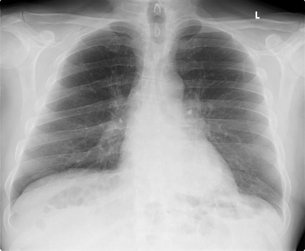
 Left Lower Lobe Pneumonia Radiology Case Radiopaedia Org
Depending on the part of the lung affected eg.

Lower lobe pneumonia. Summary Lobar Pneumonia vs Bronchopneumonia. Collapse of the left lower lobe can give rise to a double left heart border where the triangularly shaped opacity of the collapsed left lower lobe sits behind the heart and creates a second edge next to the edge of the heart. Left-sided pneumonia occurs much less frequently than the right.
According to the organism causing the infection. Pneumonia may manifest as upper abdominal pain when lower lobe infection irritates the diaphragm. Lobar pneumonia references a form of pneumonia that affects a specific lobe or lobes of the lung.
Lung Foundation Australia 2020. Videos you watch may be added to the TVs watch history and influence TV recommendations. Gastrointestinal symptoms nausea vomiting diarrhea are also common.
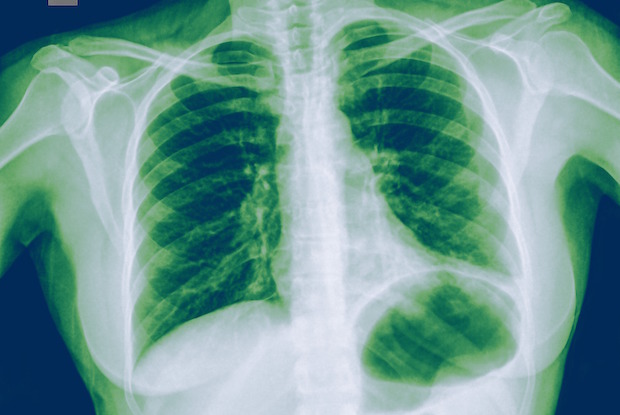
Fungal pneumonia is caused by breathing in fungal spores. Reduction of the volume of lung tissue accumulation of fluid in the pleural cavity narrowing of the lumen of the bronchi allergic reactions from other organs abscess of the lung. Left lower lobe pneumonia refers to focal diseases which is due to the presence of a virus fungus or bacteria affecting the lower respiratory tract.
March 4 left lower lobe pneumonia. This inflammation makes it. When the infection is confined to only one or few lobes of lungs that is known as lobar pneumonia.
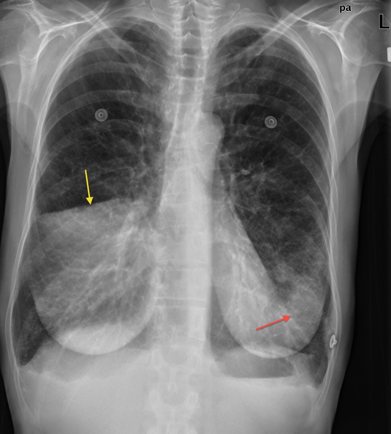
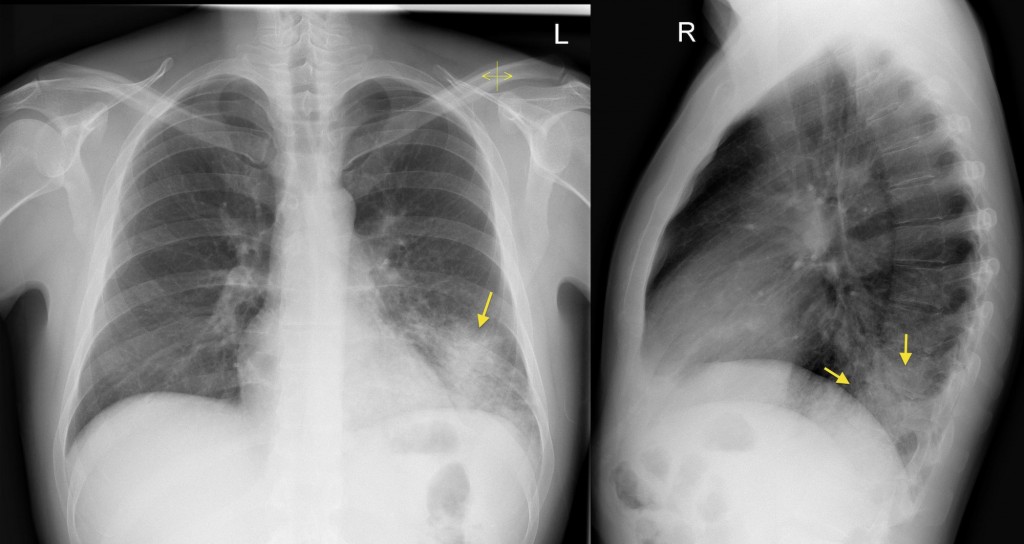
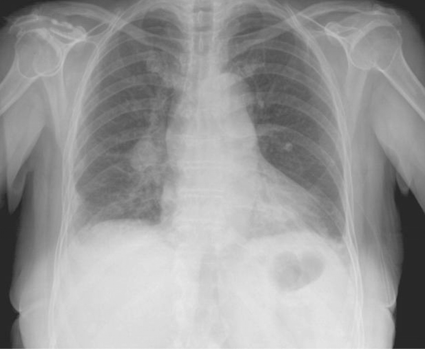
It has resolved after antibiotics34 yr old healthy female. Symptoms become variable at the extremes of age. Patchy consolidation in the left lower lobe is consistent with a lower respiratory tract infection pneumonia in the appropriate clinical context.
Lower lobe pneumonia can lead to the following complications. Right lower lobe had small fluid. A virus bacteria or fungus causes the tiny sacs of the lungs called alveoli to become inflamed and fill with fluid or pus causing a range of.
WebMD 2018 There are other ways in which pneumonia may be classified or described. Can i get this again. The antibiotic is chosen based on the causative organism identified or suspected.
The Alphabetic Index supports this advice. Bronchial pneumonia or lower lobe pneumonia. A diagnosis of lobar pneumonia pneumonia that mentions the affected lobe or multilobar pneumonia pneumonia affecting more than one lobe describes the specific site of the pneumonia rather than a type of pneumonia and would be coded according to the responsible organism if known.
This is due to the peculiar structure of the bronchial tubes. Histological lesions are always located within large zones of altered lung parenchyma which correspond to an inflammatory exudate with fibrin and some capillary congestion ensuing at the 3rd to 7th day of pneumonia. Children can present with abdominal pain as the only symptom of pneumonia.
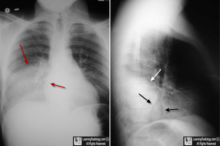
Bronchopneumonia is the inflammation of lung parenchyma that arises from bronchi or bronchioles secondary to an infectionAs given in their definitions lobar pneumonia is confined to one or few lobes but bronchopneumonia affects a wide. If playback doesnt begin shortly try restarting your device. This case illustrates the importance of assessing the lung bases when interpreting an abdominal x-ray.
This is a bacterial pneumonia and is most commonly community acquired. The infection inflames the air sacs in your lungs or the alveoli which fill with fluid or pus. In some cases this does not occur since the two can overlap sufficiently as to be indistinguishable.
These foci of pneumonia are predominantly distributed in lower lobes and dependent zones of the lungs. Double pneumonia is a lung infection that affects both of your lungs.
 The Radiological Diagnosis Of Pneumonia In Children Pneumonia Full Text
 Left Lower Lobe Pneumonia Radiology Case Radiopaedia Org
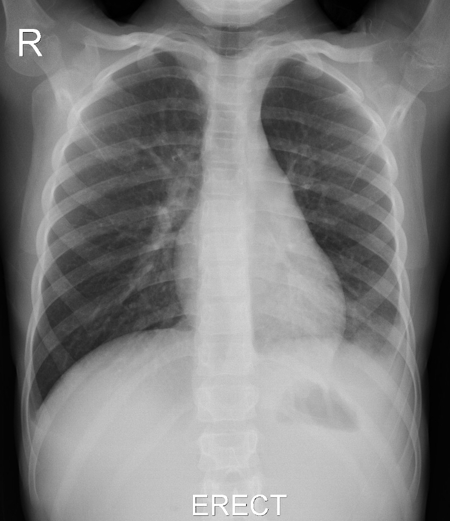
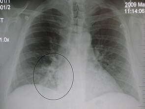
 Chest Radiography From A Patient With Right Lower Lobe Pneumonia Download Scientific Diagram
 Chest X Ray Showing Right Lower Lobe Pneumonia Download Scientific Diagram
 Pneumonia Diagnosis And Management Gponline
 Left Lower Lobe Pneumonia Radiology Case Radiopaedia Org
Chest Xray From First Presentation At 13 Weeks Showing Left Lower Lobe Download Scientific Diagram
 Community Acquired Pneumonia Pulmonary Disorders Msd Manual Professional Edition
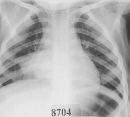
 Pneumonia Right Lower Lobe Radiology Case Radiopaedia Org Radiology Radiology Imaging Human Anatomy And Physiology
 Occult Pneumonia Wikipedia
 A Turbulent Cause Of Bilateral Pneumonia European Respiratory Society
Tidak ada komentar:
Posting Komentar
Catatan: Hanya anggota dari blog ini yang dapat mengirim komentar.